
Only nanometers in size and yet the new coronavirus SARS-CoV-2 manages to put the world on hold and change the everyday lives of many people in a way never seen before. But what are coronaviruses actually? What significance do they have for animal and human health? Which corona viruses do we already know and how does a virus suddenly manage to infect people? On closer examination of these questions, it quickly becomes clear that coronaviruses are an issue that involves the health of animals, humans and the environment.
What are coronaviruses?
Coronaviruses are viruses that occur in many different animals all over the world. They are classified in the subfamily Orthocoronaviridae (order: Nidovirales, suborder: Cornidovirineae, family: Coronaviridae), whereby this subfamily is again divided into different genera, subgenera and species [1].
Structure of a coronavirus
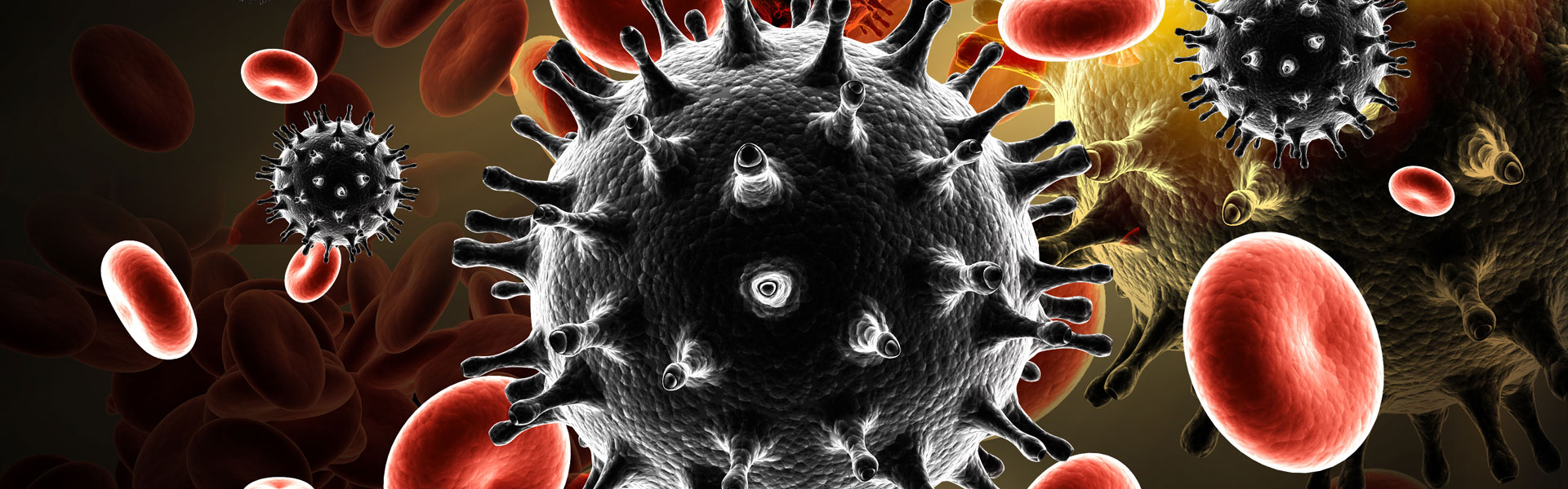
Coronaviruses have a virus envelope consisting of a lipid membrane with embedded proteins. The proteins (especially the spike protein) protruding from the membrane in coronaviruses give them their characteristic appearance under the electron microscope (see Figure 1), which led to their name (Latin corona: wreath, crown) [2]. All coronaviruses have in common that their genome, i.e. their genetic information, is in the form of a single-stranded ribonucleic acid (RNA) strand with positive polarity. This means that in coronaviruses the base sequence of the RNA corresponds to the later mRNA (messengerRNA). This has consequences for the way the virus reproduces. With a length of 26.4 - 31.7 kilobases, the corona virus genome is the longest continuous RNA genome of all known RNA viruses [3].
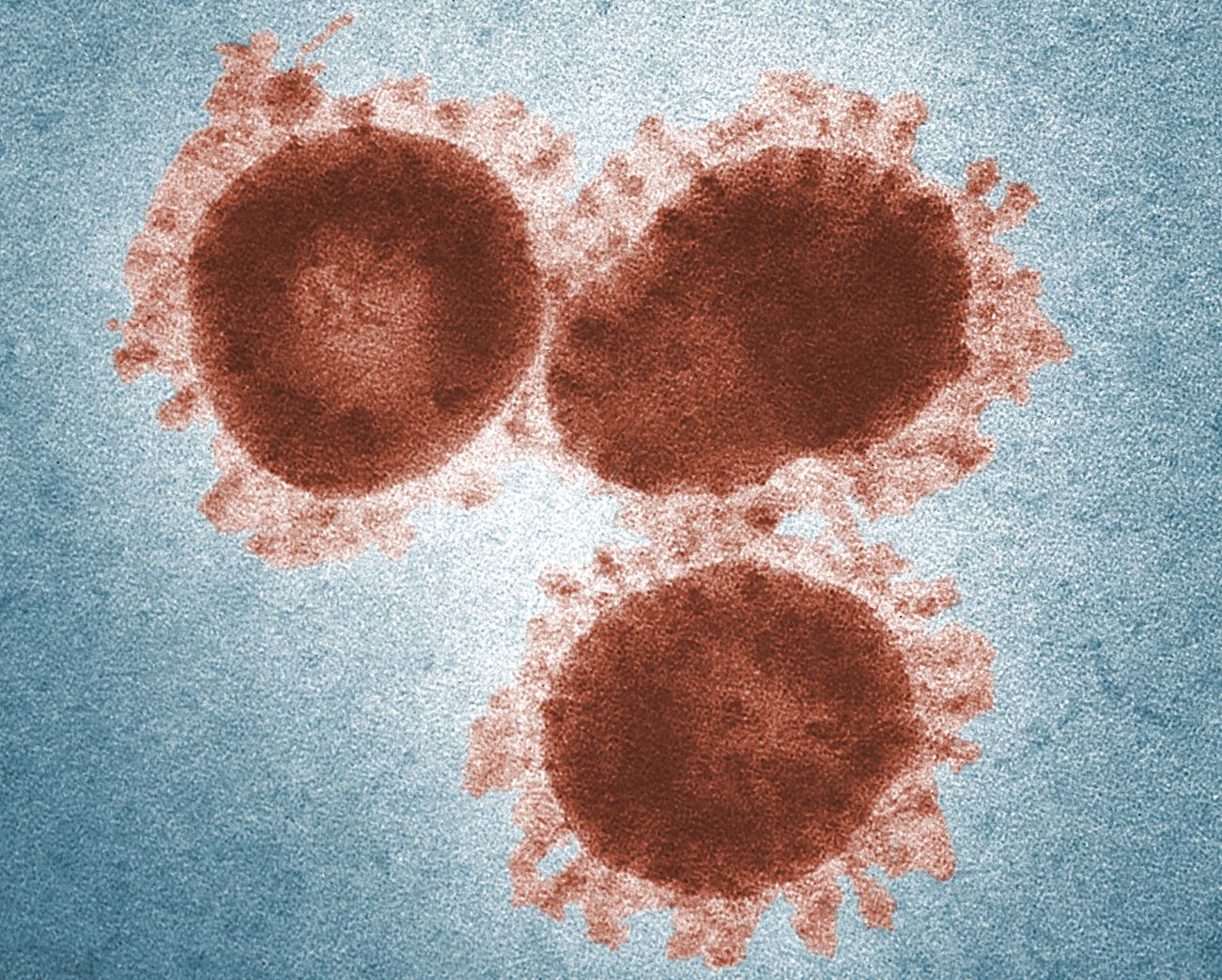
Figure 1: Corona viruses under the electron microscope. The digitally stained transmission electron microscope (TEM) image shows the avian Infectious Bronchitis Virus (IBV), which also belongs to the coronavirus family. The "wreath" around the virus particles caused by the spike glycoproteins protruding from the virus envelope has given the virus family its name. Picture: CDC/ Dr. Fred Murphy; Sylvia Whitfield
In the RNA, the information for the different viral proteins is encoded. These proteins are the nucleocapsid (N) protein surrounding the RNA genome and three membrane proteins: the spike (S) glycoprotein, the membrane (M) glycoprotein and the envelope (E) protein (48). In addition, some coronaviruses, including SARS-CoV-2, which was discovered in 2019, have a haemagglutinin (HE) on their surface (see Figure 2). The different viral proteins fulfil different tasks for the structure of the virus particles, in the reproduction of the virus, in the penetration into the host cells, in the pathogenesis and in the modelling of the immune response of the infected organism [4].

Figure 2: Schematic 3D structure of the SARS-CoV-2. The cross-section on the right shows the RNA inside the virus particle together with the nucleocapsid (N) protein. The virus is surrounded by an envelope in which various viral proteins are embedded: (spike(S) glycoprotein, membrane (M) glycoprotein, envelope (E) protein). Picture: Scientific Animations/ Creative Commons
Where do coronaviruses occur?
Coronaviruses in animals
The first coronavirus described was the Infectious Bronchitis Virus (IBV), which was isolated from chicken embryos in 1937 [5]. Since then, numerous coronaviruses have been detected in various animals, including wild animals, livestock and pets. They are divided into the genera of mammalian-associated alpha- and betacoronaviruses and bird-associated gamma- and deltacoronaviruses [6]. In veterinary medicine, different coronaviruses are of relevance. For example, the transmissible gastroenteritis coronavirus (TGEV) or the porcine epidemic diarrhea virus (PEDV) can cause severe diarrhoea in pigs. Additionally also cattle (Bovine coronavirus, BCoV), cats (Feline infectious peritonitis virus), mice (Murine coronavirus) or chickens (IBV) can be affected by coronaviruses. The spectrum of diseases caused by coronaviruses in animals ranges from mild to severe intestinal, respiratory or systemic diseases [7]. However, there are also many coronavirus infections in animals that do not seem to cause any symptoms.
Widespread in the animal kingdom
Studies on coronaviruses in a wide variety of animal species have shown that there are coronaviruses that occur in only one animal species (host-specific) and others that can infect a wide range of different animal species (host-unspecific). For example, betacoronavirus 1 was detected in cows, horses, dogs, humans, deer, antelopes, camels and giraffe. Other coronaviruses, on the other hand, only occur for example in one specific bat family. In particular, the SARS (Severe Acute Respiratory Syndrome) pandemic in 2002/2003 has led to an increase in studies on the occurrence of coronaviruses in wild animals all over the world. The greatest diversity of coronaviruses has been found in bats (summarised in [8]). However, it can be assumed that there are still some gaps in the detection of coronaviruses in wild animal populations. In particular, the data for economically and/or politically unstable regions of the world is still incomplete [8].
Coronaviruses in humans – cause of colds
The first human coronaviruses (HCoV) were described in the 1960s. These were HCoV-229E [9] and HCoV-OC43 [10]. By now, four endemic human coronaviruses are known (-229E, -OC43, -NL63, -HKU1), which circulate in the world population. In most cases, the endemic human coronaviruses cause diseases of the upper and lower respiratory tract. According to the Helmholtz Centre for Infection Research, coronaviruses are responsible for about one third of all "colds" in humans. Asymptomatic infections have also been described. In some cases, especially in immunocompromised persons, children or persons with pre-existing pulmonary diseases, acute respiratory diseases (ARE) can also occur. (Summarised in [11])
Cause of pandemics
SARS-Cov
The situation is different, however, with the human coronaviruses of zoonotic origin, which were first described in the 21st century and triggered pandemics through the transfer of animals to humans and subsequent human-to-human chains of transmission. This first time this occurred with the Severe Acute Respiratory Syndrome Coronavirus (SARS-CoV) [12]. This virus, originating in China, caused several serious respiratory diseases in humans in 2002 and 2003. About 8.000 people were affected by the disease at that time, with a mortality rate of about 9.5 %. Thanks to the rapid development of a diagnostic test and far-reaching measures to isolate those infected, the spread of the virus could be stopped. No SARS infections in humans have been reported since 2004. Later investigations in wildlife showed that SARS-related corona viruses are found in bats and civets, which is why it is assumed that the virus has spread from bats to humans via civets [13]. (see Figure 4)
MERS-CoV
Middle East respiratory syndrome coronavirus (MERS-CoV) was isolated for the first time in 2012 from a patient admitted to hospital with acute pneumonia in Saudi Arabia [14]. By 2019, approximately 2500 reported MERS-CoV infections in humans had been recorded, with approximately 30% of patients dying from the infection [11]. The main risk area for MERS-CoV infections is the Arabian Peninsula. Here, infections have been recorded both via human-to-human transmission and contact with dromedaries. Dromedary represent a reservoir for MERS-CoV [15].
SARS-CoV-2
At the end of December 2019, China reported a high incidence of pneumonia in the city of Wuhan. In January 2020, a new type of betacoronavirus was identified as the causative agent [16]. Because of its similarity to SARS-CoV, the WHO (World Health Organization) named the new coronavirus SARS-CoV-2, and the disease caused by the virus was officially named COVID-19 (Coronavirus Disease 2019). The genome of the new coronavirus shows similarities to other betacoronaviruses found in bats [16]. It can therefore be assumed that the virus originally originated in bats and, over time, has spread to other animal hosts and eventually to humans through changes. SARS-CoV-2 can be transmitted very efficiently from person to person and has therefore spread rapidly to all continents in our globalised world. In the resulting pandemic, 244,523 people were infected and 10,030 patients died (status: 20.03.2020, source: Johns Hopkins University). It is to be assumed that until a suitable active substance for the treatment of the infected persons is developed or until a suitable vaccine SARS-CoV-2 is available, SARS-CoV-2 will still be a major burden for the world population.
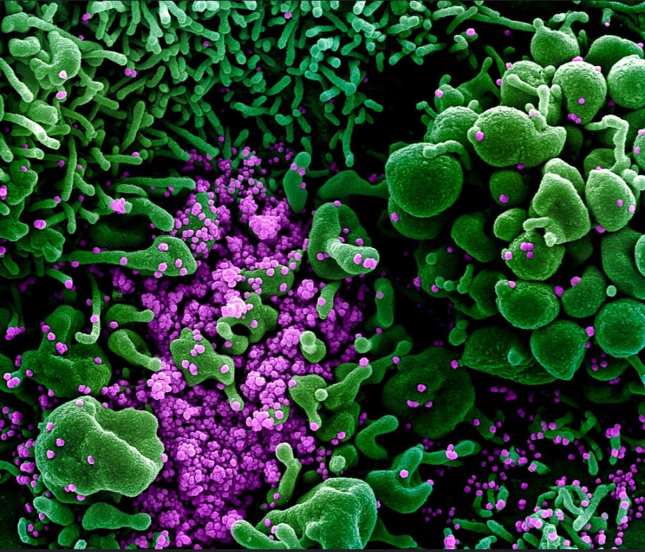
Figure 3: Coloured scanning electron microscope image of an apoptotic cell (green) infected with SARS-COV-2 virus particles (violet) and isolated from a patient sample. The image was taken at the Integrated Research Facility (IRF) of NIAID in Fort Detrick, Maryland, and colour processed. Picture: National Institute of Allergy and Infectious Diseases
Zoonoses - The leap from animals to humans
In order to infect a new host, a virus must first overcome a number of barriers. This concerns on the one hand the ability to invade the cells of the new host and multiply, and on the other hand, the ability to bypass the immune system of the host organism to such an extent that at least cell infection is possible. Since a virus cannot make directed "adaptations", these new properties are the result of random mutations (changes in the genetic material) or recombination of different coronaviruses. Coronaviruses have a high mutation rate due to their error-prone RNA polymerase, which is responsible for the duplication of genetic information [17]. In addition, homologous recombination frequently occur in coronaviruses [18]. These properties have contributed to a great diversity of coronaviruses in nature, which enables these viruses to infect numerous species. Thus, seven known coronaviruses have managed over time to transfer to humans via animal intermediate hosts (see Figure 4, modified from Corman et al. [13]). It can be assumed, that pathogenic coronaviruses from zoonotic sources will continue to spread to the human population in the future [19].
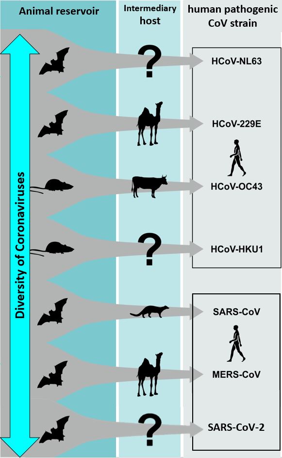
Figure 4: Virus reservoirs and potential intermediate hosts of the four endemic human coronaviruses (upper rectangle) and the three zoonotic, epidemic human coronaviruses (lower rectangle). Graphic modified from Corman et al. [13]
Research on coronaviruses as a One Health mission
The seven coronaviruses detected in humans, and in particular the zoonotic spillover events that have led to three epidemics in twenty years, demonstrate the zoonotic potential of coronaviruses. Since the viruses play a role in both veterinary and human medicine, expertise can complement each other for future research. Against the background of increasing international travel and global trade, as well as changes in climate with associated effects on pathogens and host species, presumably the risk from zoonoses will change. Important research areas for the future are in particular the research into vaccines and medicine, the expansion of knowledge about the epidemiology of coronaviruses in wild animal populations and research into prediction models for zoonotic transmissions to humans. Only by pooling expertise and by encouraging the cooperation of interdisciplinary research teams can we promote progress and success in this research field and prevent pandemics in the future.
Text: Dr Dana Thal for the German Research Platform for Zoonoses
Literature:
[1] International Comitee on Taxonomy of Viruses (ICTV), „Virus Taxonomy: 2018b Release,“ 2019.
[2] „Virology: Coronaviruses,“ Nature, Bd. 220, p. 650, 1968.
[3] P. Woo, S. Lau, Y. Huang und K.-Y. Yuen, „Coronavirus diversity, phylogeny and interspecies jumping,“ Experimental Biology and Medicine, Bd. 234, Nr. 10, pp. 1117-1127, October 2009.
[4] A. Fehr und S. Perlman, „Coronaviruses: an overview of their replication and pathogenesis,“ Methods in Molecular Biology, Nr. 1282, pp. 1-23, 2015.
[5] F. Beaudette und C. Hudson, „Cultivation of the virus of infectious bronchitis.,“ J Am Vet Med Assoc, Nr. 90, p. 51–58, 1937.
[6] P. Woo, S. Lau, C. Lam, C. Lau, A. Tsang, J. Lau, R. Bai, J. Teng, C. Tsang, M. Wang, B. Zheng, K. Chan und K. Yuen, „Discovery of seven novel Mammalian and avian coronaviruses in the genus deltacoronavirus supports bat coronaviruses as the gene source of alphacoronaviruses ans betacoronaviruses and avian coronaviruses as the gene source of gamma- and deltacoronavirus,“ Journal of Virology, Bd. 86, pp. 3395-4008, 2012.
[7] L. Saif, „Animal coronaviruses: What can they teach us about the severe acute respiratory syndrome?,“ OIE Revue Scientifique et Technique, Bd. 23, Nr. 2, pp. 643-660, 2004.
[8] J. F. Drexler, V. M. Corman und C. Drosten, „Ecology, evolution and classification of bat coronaviruses in the aftermath of SARS,“ Antiviral Research, Bd. 101, pp. 45-56, January 2014.
[9] D. Hamre und J. Procknow, „A New Virus Isolated from the Human Respiratory Tract,“ Proceedings of the Society for Experimental Biology and Medicine, Bd. 121, Nr. 1, pp. 190-193, January 1966.
[10] K. McIntosh, J. Dees, W. Becker, A. Kapikian und R. Chanock, „Recovery in tracheal organ cultures of novel viruses from patients with respiratory disease,“ PNAS, Bd. 57, Nr. 4, pp. 933-940, April 1967.
[11] V. Corman, J. Lienau und M. Witzenrath, „Coronaviren als Ursache respiratorischer Infektionen,“ Internist, Nr. 60, pp. 1136-1145, 2019.
[12] C. Drosten, S. Günther, W. Preiser, S. van der Werf, H.-R. Brodt, S. Becker, H. Rabenau, M. Panning, L. Kolesnikova, R. A. Fouchier, A. Berger, A.-M. Burguière und e. al., „Identification of a Novel Coronavirus in Patients with Severe Acute Respiratory Syndrome,“ New England Journal of Medicine, Nr. 348, pp. 1967-1976, 2003.
[13] V. M. Corman, D. Muth, D. Niemeyer und C. Drosten, „Chapter Eight - Hosts and Sources of Endemic Human Coronaviruses,“ Advances in Virus Research, Nr. 100, pp. 163-188, 2018.
[14] A. M. Zaki, S. van Boheemen, T. M. Bestebroer, A. D. Osterhaus und R. A. Fouchier, „Isolation of a Novel Coronavirus from a Man with Pneumonia in Saudi Arabia,“ New England Journal of Medicine, Nr. 367, pp. 1814-1820, November 2012.
[15] I. M. Mackay und K. E. Arden, „MERS coronavirus: diagnostics, epidemiology and transmission,“ Virology Journal, Bd. 12, Nr. 222, 2015.
[16] N. Zhu, D. Zhang, W. Wang, X. Li, B. Yang, J. Song, X. Zhao, B. Huang, W. Shi, R. Lu, P. Niu, F. Zhan und et al., „A Novel Coronavirus from Patients with Pneumonia in China, 2019,“ New England Journal of Medicine, Nr. 382, pp. 727-733, February 2020.
[17] S. Duffy, L. A. Shackelton und E. C. Holmes, „Rates of evolutionary change in viruses: patterns and determinants,“ Nature Reviews Genetics, Nr. 9, p. 267–276, March 2008.
[18] M. M. Lai, „RNA Recombination in Animal and Plant Viruses,“ Microbiological Reviews, Bd. 56, Nr. 1, pp. 61-79, March 1992.
[19] C. M. Coleman und M. B. Frieman, „Coronaviruses: Important Emerging Human Pathogens,“ Journal of Virology, Bd. 88, Nr. 10, p. 5209–5212, May 2014.